The first description of histopathology of Lates calcarifer herpesvirus (LCHV) infection in barramundi (Lates calcarifer)
26/05/2023Barramundi (Lates calcarifer), an economically important species for mariculture in the Asia-Pacific region, has been threatened by various infectious diseases. Lates calcarifer herpes virus (LCHV) is a newly emerging virus which was first reported in farmed barramundi in 2015.
Up to now, the pathognomonic histopathological lesion of this disease has not yet been described for the purpose of disease diagnosis. This study investigated the histology of five natural LCHV outbreaks throughout a year. Clinical signs of barramundi naturally infected with LCHV showed dark body with scale loss, eroded and necrotic tail (heavy infected fish with more than 80% scale loss), opaque eye and the liver was swelling with white patches (Figure 1). In particular, consistently detected the presence of a typical type of intra-nuclear inclusion bodies (IIBs) in various organs of infected fish (Figure 2). Experimental infection was conducted by injecting the healthy fish with filtered supernatant of crude extract from the LCHV-positive barramundi from the field. Histological examination of experimentally infected barramundi clearly indicated the presence of the same type of IIBs in multiple organs, including the liver, pancreas, kidney, eyes, gills, and adipose tissue (Figure 3).
The presence of IIBs in barramundi infected with LCHV was a pathognomonic feature that can be used for the histological diagnosis and particularly for surveillance of this emerging viral disease in barramundi farming countries.
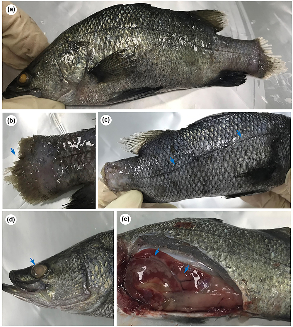
Figure 1. Clinical signs of barramundi naturally infected with LCHV: (a) dark body with scale loss; (b) eroded and necrotic tail (arrow); (c) heavy infected fish with more than 80% scale loss (arrow) and an eroded tail; (d) opaque eye (arrow); (e) the liver was swelling with white patches (arrows)
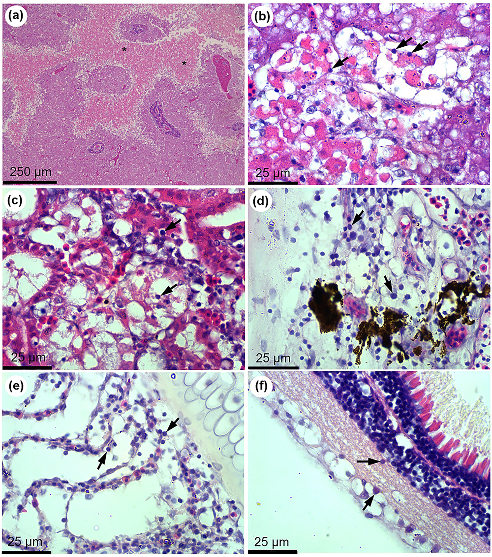
Figure 2. Histopathology of naturally LCHV infected barramundi. (a) multifocal necrosis in the liver; (b) Intra-nuclear inclusion bodies (IIBs, arrows) were present in multiple necrotic areas of the liver; (c) IIBs (arrows) in degenerating tubules and haematopoietic tissue of the kidney; (d) multiple IIBs (arrows) in the epithelium of the skin; (e) presence of IIBs (arrows) inside gill filaments; (f) retina of cloudy eye contained IIBs (arrows)
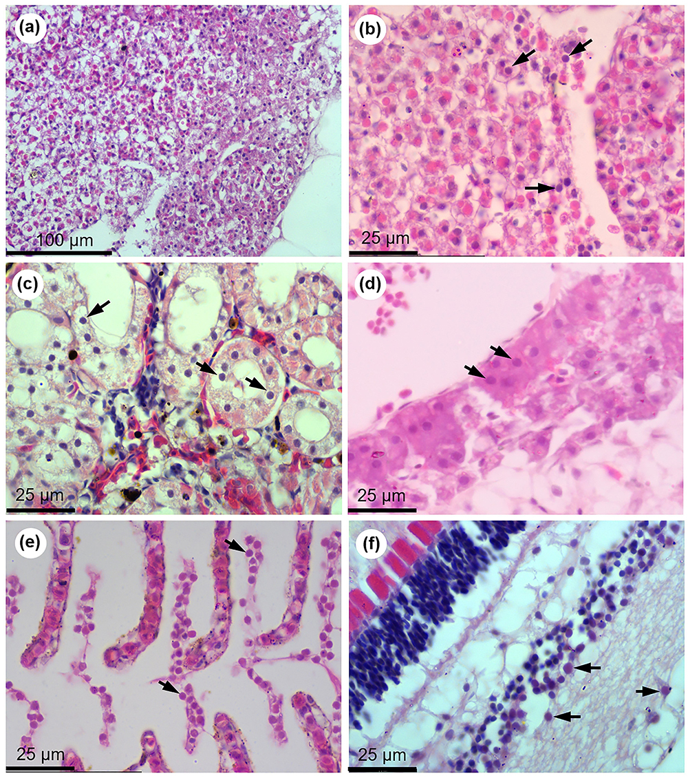
Figure 3. Presence of intra-nuclear inclusion bodies (IIBs, arrows) in multiple organs including the liver (a, b), kidney (c), pancreas (d), gills (e) and retina of the cloudy eye of barramundi infected with LCHV in the experimental condition.
Source: https://doi.org/10.1016/j.aquaculture.2022.739091;
Link: https://www.sciencedirect.com/science/article/abs/pii/S004484862201208X
Tran Duc Dien (Coastal Branch - VRTC)